If you've ever used your phone's camera to see a digital arrow floating over a real street, you already understand the basic idea behind a new surgical tool. Researchers have built an augmented reality (AR) system that overlays a 3D picture of a tumor onto the live video a surgeon sees during keyhole surgery. Here is what it does, how it works, and why it matters beyond the operating room.
The work was reported by Medicalxpress. The details below come from that report, and it does not specify device models or software versions, so none are listed here.
The problem: tumors you can't see
In laparoscopic, or keyhole, surgery, the surgeon works through small incisions and watches a camera feed on a screen. According to Medicalxpress, tumors beneath the surface of the liver can be difficult to locate this way. The organ looks like a smooth surface on screen, while the target sits out of sight underneath.
That is the gap the new technology tries to close.
What the system does: GPS inside the organ
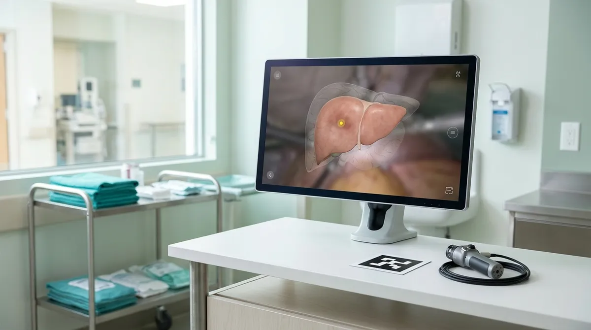
The system uses augmented reality to overlay a 3D visualization of the tumor and other anatomy onto the surgeon's laparoscopic camera view. Think of it as a navigation layer. Your car's GPS puts the route on top of the real road; this puts the hidden tumor on top of the real liver.
The surgeon keeps looking at the same camera image. The difference is that the buried structures are now drawn into it.
How a QR-code marker guides it
Per the report, a QR-code marker guides the system. You've likely scanned one to open a website, and the principle is similar here: a QR code is easy for a camera to recognize and track. A marker like this gives software a fixed reference point, so it can line up the digital 3D model with what the camera is actually seeing.
The source summary does not detail exactly how the marker is placed or calibrated, so we won't guess at the specifics. The key idea is simple: a recognizable reference point lets the overlay stay anchored to the real anatomy.
First patient use and what comes next
The technology has now been used in its first patient. Medicalxpress reports that the team plans to validate the system and expand its use, and that more liver operations are planned. That means it is early-stage: one clinical use is a milestone, not proof that it works reliably across many cases. Validation is the step that answers that question.
Surgeons and mathematicians, working together
The project is also a good example of interdisciplinary problem solving. The coverage describes surgeons and mathematicians collaborating on a practical operating-room problem. Surgeons know what they can't see; mathematicians know how to build and align 3D models. Neither could solve this alone.
What this means for everyday tech
You won't be using this at home, but it shows where common tools are heading:
- Cameras as sensors. The same kind of camera and marker tracking used in consumer AR apps is being applied to high-stakes work.
- Overlays instead of guesswork. Putting hidden information directly on a live view is more useful than flipping between screens.
- Simple markers, big payoff. A QR code is a low-tech anchor that makes complex software more dependable.
If you want to experience the consumer version, try the measuring or object-placement features built into your phone's AR tools. Point the camera, watch digital content lock onto a real surface, and you'll see the same principle that this surgical system applies to a liver.
The takeaway
When you hear about AR in medicine, look for the practical question it answers: what was hidden, and how does the system keep the overlay lined up with reality? Here, the answer is a 3D tumor map anchored by a QR-code marker. Watch for the validation results, because they will show whether this early success becomes routine care.