You have probably used augmented reality to preview a sofa in your living room or catch a creature on your phone screen. The same basic idea is now being used in an operating room, and understanding how it works says a lot about where everyday AR technology is heading.
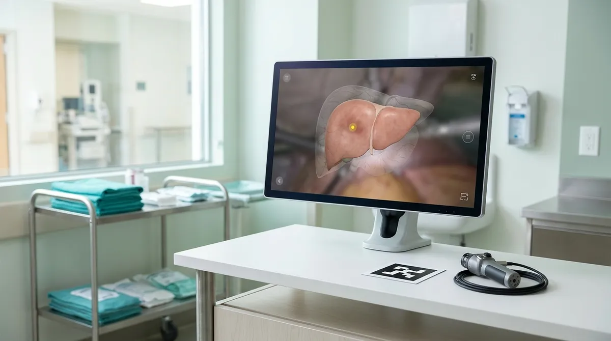
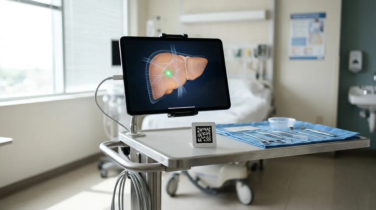
Per a report from Medicalxpress, a new system uses augmented reality to overlay a 3D visualization of a tumor and other anatomy onto the live camera view during laparoscopic (keyhole) surgery. The target is liver tumors that sit beneath the organ's surface and are hard to locate. According to the report, the system has now been used on its first patient.
Below is a plain-language look at the problem, the approach, and what to watch next. I'm limited to what has been publicly reported, and the report leaves out some details, such as the specific hardware and software versions involved, so I won't guess at them.
The problem: a tumor you can't see
In keyhole surgery, the surgeon works through small openings and watches a camera feed on a screen. That feed shows the surface of the organ. Per the report, tumors beneath the liver's surface can be difficult to locate this way.
Think of it like trying to find a stud behind drywall by looking only at the paint. You know something is there, but the surface doesn't show you where.
The idea: a map laid over the live view
The system's answer is to place a 3D visualization of the tumor and other relevant structures directly over the surgeon's camera view. Instead of mentally matching a scan to what's on screen, the surgeon sees the hidden structures displayed in context.
A helpful comparison is the navigation app on your phone. A paper map shows the whole city, but a GPS dot shows where you are on it right now. The "GPS inside the organ" comparison is my framing, not a claim from the researchers, but it captures the goal: turning a static picture into live guidance.
The anchor: a QR-code marker
For an overlay to be useful, the software has to know where the organ is and how it's oriented. According to the report, a QR-code marker guides the system.
You already know why this works if you've ever scanned a code with your phone camera. A QR code is an easy-to-recognize pattern, so software can quickly figure out where it is and which way it's facing. Using a marker like that gives the system a fixed reference point to line up the 3D model with the live image.
The report as summarized here doesn't spell out every technical step of how the alignment is done, so treat this as the general principle rather than a full description of the method.
What has happened so far
The key milestone is that the technology has been used in a real operation on a first patient. That moves it from a lab concept toward clinical practice. Per the report, there are plans to validate the technology and expand its use, with more liver operations planned.
That word "validate" matters. One successful use is an encouraging start, not proof that the system is better than current methods. Larger testing is what will show how accurate and useful it is.
Why the team behind it is interesting
The angle that stands out to me is the collaboration. The report frames this as a practical operating-room problem tackled by surgeons together with mathematicians. Surgeons know what they need to see and when. Mathematicians can build the models and methods that make a flat camera image and a 3D scan agree with each other.
That pairing is a pattern worth noticing in any field: the best tools often come from people who understand the problem and people who understand the math, working in the same room.
What this means for everyday tech users
You won't be buying this at an electronics store, but the building blocks should look familiar:
- A camera feed. The same kind of live video your phone uses.
- A recognizable marker. A QR-style code that tells software where things are.
- A 3D overlay. Digital information placed on top of the real world.
When you see AR on your phone, you're using a simpler version of this recipe. Seeing it applied to surgery shows how far the approach can go when accuracy matters.
What to watch for next
If you follow this area, here are the questions that will tell you whether the technology is maturing:
- Does validation hold up? Look for results from more patients, not just the first.
- Does it expand? The plan is to use it in more liver operations. Whether it spreads to other organs or procedures is an open question.
- Does it become routine? The bigger shift would be real-time navigation becoming a normal part of keyhole surgery rather than a special case.
None of these are guaranteed, and the report doesn't promise them. They're simply the milestones that would separate a promising first case from a lasting tool.
The takeaway
Next time an AR app places a digital object into your room, remember the principle behind it: a camera, a marker, and a model that lines up with the real world. Then follow the validation results for this liver-surgery system, because that is where we'll learn whether "GPS inside the body" is a milestone or just a first step.